Research

A summary of the ongoing research projects in the lab

Our mission

The goal of the Krause Laboratory is to define how stem and progenitor cells balance and regulate fate decisions in health and disease. We study stem cells to improve strategies for repair and regeneration of the hematopoietic system as well as solid organs including the lung and parathyroids.


Diane discusses how the Krause Lab leverages the interface of fundamental science and clinical medicine at Yale to understand stem cell biology and improve patients’ lives.

Hematopoietic cell self-renewal and differentiation

Using bone marrow derived stem and progenitor cells to understand blood development

Watch cell fate commitment unfold as a single bipotent Megakaryocytic-Erythroid Progenitor differentiates into megakaryocytes (green) and erythroid cells (red).

“What are the molecular mechanisms that regulate the development of healthy and cancerous blood cells?”

Using state of the art approaches including CRISPR, genetic screens, and timelapse microscopy, the Krause Lab works to identify the molecular mechanisms that regulate hematopoiesis. One major focus is determining how progenitors pick which differentiated cell type to become – this is cell fate commitment. Cell fate decisions are regulated by differential transcription and epigenetics and the cell cycle, in ways that are not yet understood. These processes affects the decision of the bipotent megakaryocyte-erythrocyte progenitor (MEP) to pick the megakaryocytic versus erythroid lineage.
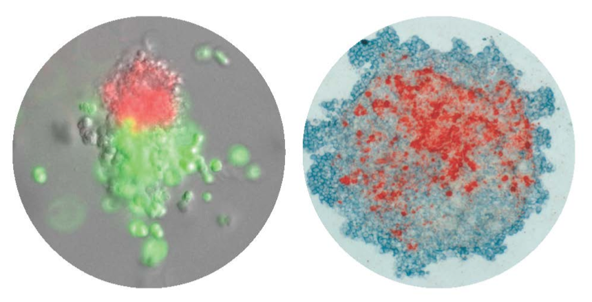
CFU-Mk/E: Colonies derived from a single cell that differentiated down the megakaryocytic and erythroid lineages. Left: Immunofluorescence, Right: Immunohistochemistry.

CFU-Mk/E: Colonies derived from a single cell that differentiated down the megakaryocytic and erythroid lineages. Left: Immunofluorescence, Right: Immunohistochemistry.

We have pioneered techniques for time-lapse imaging of the dynamics of differentiation in individual MEP (see video). The Krause lab has also developed immunohistochemistry and immunofluorescence methods that aid in high-throughput characterization of differentiation markers (see figure). In our ongoing research, we are applying cutting edge genomic approaches like single cell RNA-sequencing to finely study transcriptomic changes during the dynamic processes of hematopoiesis and leukemogenesis.
Megakaryopoiesis

Transcriptional and Epigenetic control of megakaryocyte maturation

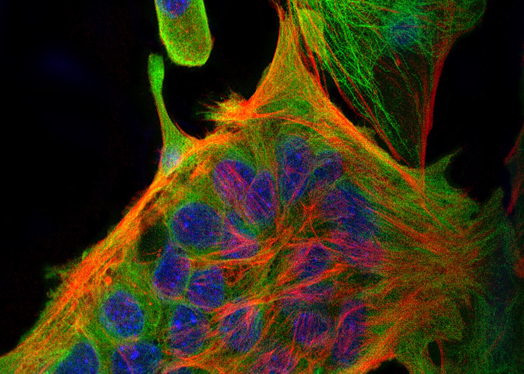
Primary megakaryocyte stained for cytoskeleton (actin and tubulin) and DNA (DAPI, blue).

“Megakaryocytes are truly amazing cells.”

Megakaryocytes (Mk) are truly one of the most fascinating cell types in the body. After a 2N cell commits to the Mk lineage, it undergoes polyploidization (replication of its nuclear DNA without cell division) and then undergoes tremendous cytoskeletal rearrangements to become amongst the largest cells in the body jam packed with membranes and actin cytoskeletal components that then undergo proplatelet formation followed by release of about ~1000 functional platelets per Mk. Due to the physiological need for platelets for blood clotting, the average adult produces over 100 million megakaryocytes and releases over 10e11 platelets per day (that’s over 1 million platelets per second)! The Krause lab is elucidating the transcriptional mechanisms of megakaryocyte maturation with a focus on the role of the cytoskeleton, MKL1 and other transcription factors.

Leukemogenesis

Studying the genetic dysregulation that causes Acute Megakaryoblastic Leukemia

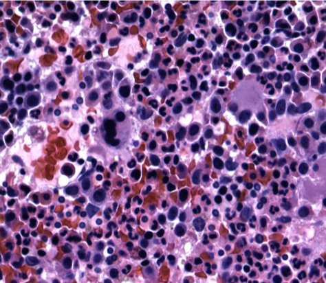
Acute Megakaryoblastic Leukemia is characterized by increased blasts in the bone marrow, seen above.

“How does improper development of megakaryocytes lead to leukemia?”

Improper development of megakaryocytes leads to Acute Megakaryoblastic Leukemia (AMKL), a deadly variant of Acute Myeloblastic Leukemia (AML) characterized by increased blasts in the marrrow. To better understand the healthy and cancerous development of these cells, we focus primarily on two genes, RBM15 and MKL1, that are fused in the t(1;22) translocation often observed in AMKL patients.

To study RBM15 and MKL1, we use patient-derived cells, murine and human embryonic stem cells, and a broad array of methods. Ongoing work on RBM15 is focused on m6A methylation, RNA stability, transcription and notch signaling. MKL1, also part of the t(1;22) translocation specific to acute megakaryoblastic leukemia, activates the transcription factor serum response factor (SRF). Ongoing work on MKL1 is focused on the mechanisms by which MKL1 promotes megakaryocytopoiesis.

Our goal is that by better understanding these two genes, we will learn more about the carefully orchestrated process of blood cell development and the ways this process breaks down in AMKL.

Parathyroid Differentiation and Function

Using stem cells to restore parathyroid function

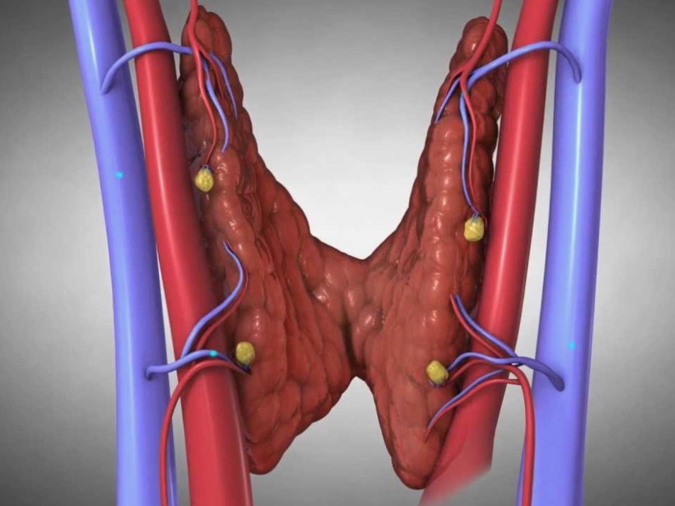
The parathyroid glands (yellow) secrete PTH and are crucial for calcium homeostasis. Image by Norman Parathyroid Center

“How can we generate functional parathyroid tissue in the laboratory to cure hypoparathyroidism?”

The parathyroid glands are small (the size of a grain of rice) tissues located adjacent to the thyroid gland in the neck. Patients with hypoparathyroidism lack parathyroid gland function. Without these glands and the hormone they produce, called parathyroid hormone (PTH), levels of calcium in the bloodstream drop. This calcium deficiency can lead to serious health problems, including painful muscle spasms in the hands and feet, seizures, an irregular heartbeat, or even heart failure. Patients also suffer on a daily basis due to fatigue and ‘brain fog,’ a less well understood aspect of the disease. Current treatments require regular calcium supplements that can be short-acting and trigger unwanted side effects, making it difficult for patients to sustain a proper balance.

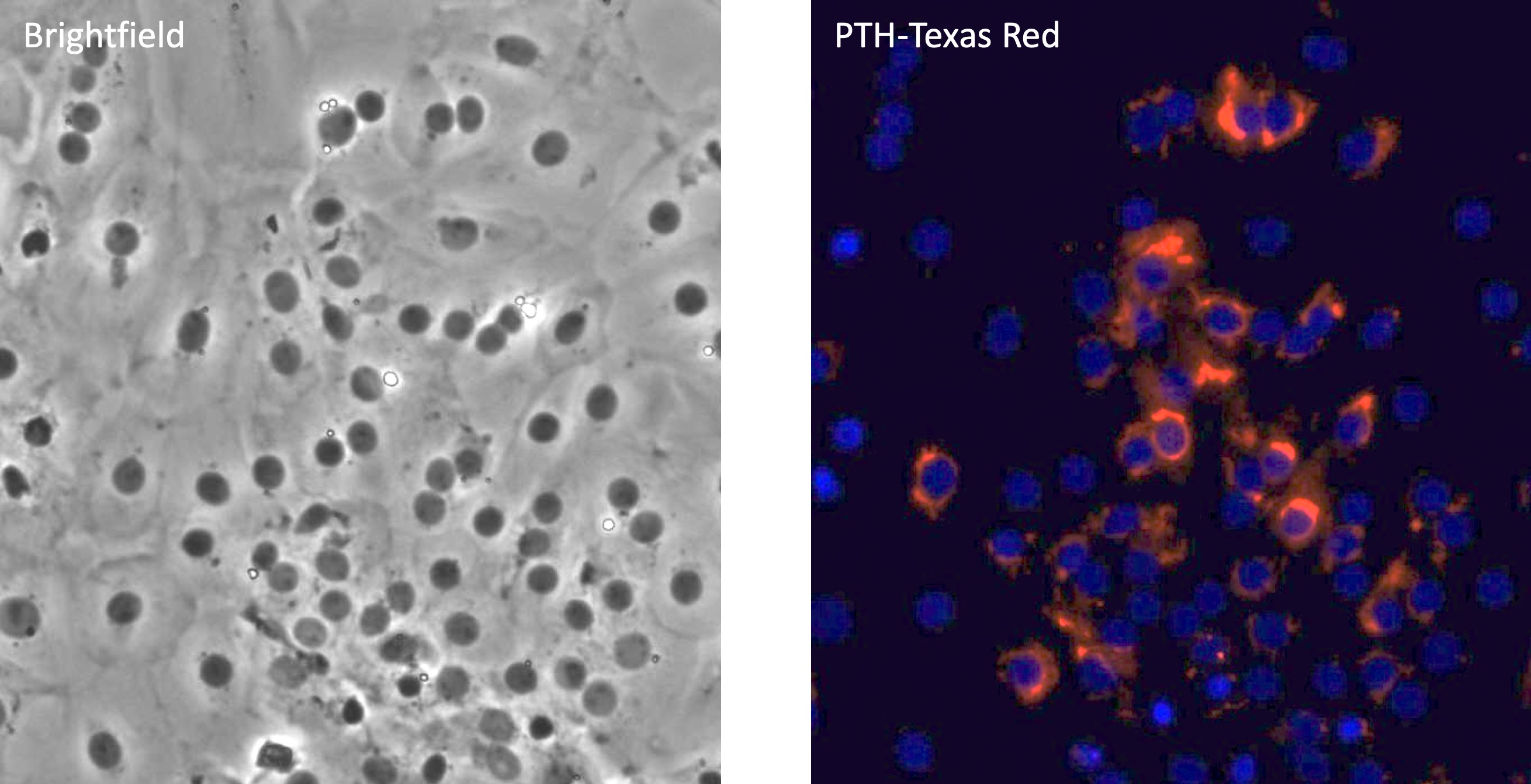
Staining of primary parathyroid cells after 4 hours of low calcium stimulation. Same field is shown on left (brightfield) and right (immunofluorescence for PTH in red, DAPI in blue). Images by Betty R. Lawton Ph.D.

Staining of primary parathyroid cells after 4 hours of low calcium stimulation. Same field is shown on left (brightfield) and right (immunofluorescence for PTH in red, DAPI in blue). Images by Betty R. Lawton Ph.D.

The most common cause of hypoparathyroidism is that these glands are removed during surgery for thyroid cancer. To be sure that no malignant tissue remains, such surgery often involves removing the parathyroid gland. Other causes of hypoparathyroidism include autoimmune diseases and hereditary conditions that lead to depleted levels of PTH.

The Krause laboratory along with collaborators across the country has taken the first steps toward transforming stem cells into parathyroid cells that can serve the same function as intact parathyroid glands. Building on earlier published efforts, the team has discovered that the completed process will need to encompass five stages of specialization from an undifferentiated stem cell to a fully functioning parathyroid-like cell. Each step involves inducing the cell to express the specific genes necessary to guide the process in the desired direction.

Advanced Cell Therapy Laboratory

Producing cell therapy products for the Yale New Haven Health system

To support clinical investigation in the field of cellular therapy the ACT Laboratory is located directly adjacent to the Transfusion Medicine Division and the Clinical Blood Bank for Yale New Haven Hospital.

“Providing world-class facilities and research support for cellular therapies.”

The Yale Advanced Cell Therapy (ACT) laboratory is a state-of-the-art laboratory designed for the production of high quality, safe, cell products for clinical treatment. The ACT is dedicated to and responsible for the production of cellular products to be used in phase I/II clinical trials conducted by investigators at Yale and in multicenter trials. Our laboratory has significant experience with IND-enabling studies using Good Manufacturing Practices. Cell product safety and quality are the top priorities for our laboratory. The ACT assures that all government regulations are met, that all products meet defined release criteria, and that all products are safe for administration to patients.

All steps of manufacturing are controlled by Standard Operating Procedures, overseen by QA/QC, and performed in our facility by qualified personnel. Cells for clinical research are isolated, expanded, tested, certified and released using validated best practices. To support clinical investigation in the field of cellular therapy the ACT Laboratory is located directly adjacent to the Transfusion Medicine Division and the Clinical Blood Bank for Yale New Haven Hospital. Click here to learn more about the GMP Facility.